Key Takeaways
-
Both genetics and hormones play important roles in lipedema development and progression. Consider family history and life-stage hormonal changes when evaluating risk.
-
Hormonal factors are why the condition often begins during puberty or pregnancy.
-
Differentiate lipedema from common obesity and lymphedema with clinical history, symptom patterns, and appropriate imaging for diagnosis.
-
Lifestyle and environmental variables including diet, exercise, and weight management can influence disease course and need to be incorporated into treatment plans.
-
Treat lymphatic function, vascular fragility, and inflammation in conjunction with genetics and hormones for a truly comprehensive treatment plan.
-
Actionable steps are to document family history and symptom onset, observe changes during hormonal events, seek focused diagnostics, and embrace an anti-inflammatory diet, regular low-impact exercise, and lymphatic-support therapies.
Lipedema is caused by a combination of hormonal and genetic factors. The condition frequently presents around the time of puberty, pregnancy, or menopause, indicating that hormones are a key factor.
Family studies and gene links indicate inherited risk that runs in families. Studies indicate that both can interplay, altering fat accumulation and triggering discomfort and edema in legs and arms.
The main body will examine existing evidence for hormones, genetics, and their intersection.
The Core Debate
Lipedema is a chronic condition characterized by bilateral, frequently asymmetric accumulation of subcutaneous adipose tissue that causes pain, swelling, and functional impairment. The crux of the debate is whether it’s hormones, genetics, or a combination of both. Below are deep dives into genetic risk, hormonal triggers, their overlap, and environmental modifiers to unpack what we do know and what we’re still uncertain of.
1. Genetic Predisposition
Family patterns run deep. With up to 60% of lipedema patients reporting a blood relative with similar symptoms, research confirms a clear hereditary component. A 2023 study of 1,091 Spanish patients substantiated this familial trend. Others suggest 60–80% of diagnosed patients have a direct family member with compatible symptoms.
That backs inherited risk; however, the precise mechanisms differ and paternal connections have been proposed in small studies. Known markers stay scant. A small number of rare variants linked to fat regulation and lymphatic development have been identified, but no monogenic cause has been established.
Genetic mutations can alter adipocyte activity, resulting in hypertrophic and hyperplastic fat cells and increasing intercellular fibrosis and macrophage infiltration in biopsies. Those tissue changes connect to chronic inflammation and pain.
Distinction matters: nonsyndromic primary lipedema presents mainly as isolated adipose expansion, while syndromic forms overlap with other connective tissue or lymphatic disorders and often follow different inheritance patterns.
2. Hormonal Triggers
Hormonal shifts often time the onset. Puberty, pregnancy, menopause – these are all common windows when lipedema makes its debut or exacerbates. Estrogen signaling is probably involved. Altered estrogen receptor activity can direct the location of subcutaneous fat deposition.
Steroid hormone changes affect adipogenesis, the process that creates and enlarges fat cells. Therefore, female hormones may promote fat growth in thighs, hips, and arms characteristic of lipedema. Clinical notes additionally record symptom changes with hormonal contraceptives or estrogen therapy, at times exacerbating swelling or fat accumulation and other times with no obvious impact.
Mechanisms range from changes in local blood flow and lymphatic pressure to receptor-mediated fat metabolism, but direct chains of causality are still being researched.
3. The Intersection
Genetic susceptibility and hormonal milieu join forces. Those with the predisposition have aberrant adipocyte reactions to hormonal fluctuations. There’s a lot of evidence that both inherited and hormonal factors change lipid handling, inflammation, and lymphatic stress in affected tissue.
Gene-hormone interactions may modify estrogen receptor distribution and PPARγ activity, which alters fat cells’ propensity to store lipid as well as their responsiveness to signals. This unified model accounts for variable onset, asymmetry, and progressive stages more effectively than a one-cause perspective.
4. Environmental Influence
Diet, activity, and weight management change course. Good nutrition and daily exercise control edema and promote lymphatic health. They seldom reverse primary fat alterations.
Environmental cues can exacerbate inflammation and weight gain in genetically susceptible individuals. Practical steps include an anti-inflammatory diet, low-impact exercise, compression, and lymphatic care to improve outcomes.
Hormonal Influence
Hormones are key players in determining adipose tissue mass and fat distribution. Estrogen, progesterone, and androgens all influence lipid uptake, storage, and breakdown in adipocytes. Estrogen promotes subcutaneous fat growth, particularly in the lower body, while androgens prefer visceral depots.
Progesterone has modulatory effects on both adipocyte metabolism and fluid balance that can transform tissue swelling. These hormones signal through receptors and local enzyme systems in adipose tissue, altering gene expression and cell behavior in ways that can promote lipedemic patterns.
Puberty
Puberty causes a sharp increase in estrogen in females, which fuels the growth of subcutaneous fat in the hips, thighs, and buttocks. This is a common time for lipedema to make its initial appearance as estrogen redirects fat deposition to lower body depots and alters adipocyte metabolism.
Hormonal surges can encourage reactive over-swelling, development of small fat nodules, and the symmetry characteristic of early lipedema enlargement. Following the onset of symptoms during puberty, timing, rapidity, and pain of fat changes distinguish early lipedema from normal pubertal fat gain and pave the way for earlier and more accurate diagnosis.
Pregnancy
Pregnancy induces persistent elevations of estrogen and progesterone and increases aromatase activity, which converts androgens to estrogens locally in adipose tissue. These shifts amplify fat accumulation in hips, thighs, and legs and can exacerbate preexisting lipedema or bring out a dormant susceptibility.
Fluid retention, leaking blood vessels, and pressure on lymphatic drainage during pregnancy cause edema and tissues to feel heavier and spongier. Management is more difficult in pregnancy because treatments such as liposuction are not suitable, so conservative measures such as compression, gentle exercise, and close tracking come into play.
Look out for new or worsening symptoms during pregnancy and in the postpartum months for prompt treatment.
Menopause
Falling estrogen at menopause shifts fat from gluteal and femoral depots toward abdominal stores. For people with lipedema, this hormone loss can still induce symptom flare-ups and exacerbated edema.
Now, shifts in estrogen receptor signaling and steroidogenic enzyme balance may make fat more fragile and prone to fluid accumulation, putting postmenopausal women with lipedema at greater risk of pain and bruising. Increased ERα/ERβ ratios in lower-body adipocytes have been observed in lipedema and could alter tissue response to waning estrogen.
Treatment options for menopausal patients include:
-
Hormone replacement therapy after individualized risk assessment
-
Targeted compression and manual lymph drainage
-
Weight management and tailored resistance exercise
-
Pharmacologic support for edema and vascular health
Genetic Blueprint
Your genetic blueprint determines who stores fat where. This is key to lipedema risk. Studies consistently show a strong familial link. Between 60% and 89% of people with lipedema report affected relatives, and about 15% have a family member currently known to have the condition.
This evidence indicates a genetic disposition that changes fat metabolism and causes symmetrical, disproportionate fat deposition to become more likely in the extremities than the torso.
Family Links
Families with this disorder frequently exhibit intergenerational patterns of symmetric fat deposition on hips, thighs, and calves with first-degree relatives noted to be the most commonly affected. Reports describe clear clustering: daughters, mothers, and sisters can display similar stages of limb fat enlargement and tenderness.
This repeat pattern undergirds the practice of leveraging family history as a clinical trigger for early screening and surveillance. Gathering a basic family symptom matrix assists clinicians in identifying danger.
A handy chart could document family members, age of onset, location on the body, tenderness or bruising, and suspected lipedema stage. With that table, clinicians can stratify who requires imaging, closer follow-up, or referral for specialist care.
Early recognition based on family links can guide preventive steps: weight management for overall health, compression to reduce symptoms, and prompt referral when disproportion becomes apparent. Family awareness reduces time to diagnosis, which is commonly postponed for years.
Gene Research
Molecular studies are starting to identify pathways that may drive lipedema. Variants implicated in adipocyte function, lipid storage, and hormone signaling appear repeatedly. PPARγ-related pathways, which govern fat cell differentiation, and estrogen receptor signaling, which links to female-biased onset and shifts at puberty or pregnancy, are two prominent pathways being investigated.
AKR1C1 popped as the initial mutated gene associated with nonsyndromic familial lipedema in a single family, highlighting enzymes that convert steroid hormones as a possible mechanism. Genome-wide association and sequencing studies implicate rare variants.
One comprehensive study identified 97% of pathogenic variants with extremely low allele frequencies of less than 0.0001, emphasizing the contribution of rare, family-specific mutations. Studies with bigger family groups are required to chart the entire genetic blueprint.
GWAS can identify common risk loci, while whole-exome or genome sequencing in affected pedigrees can identify rare causal variants. These collective efforts could potentially enable genetic tests that assist in classifying lipedema subtypes and forecasting its progression.
Translating gene findings into care could enable personalized treatment by targeting PPARγ-driven pathways, modulating estrogen effects, or using gene-informed surgical planning. Ongoing studies try to connect certain markers with symptom patterns and treatment response.
Beyond The Cause
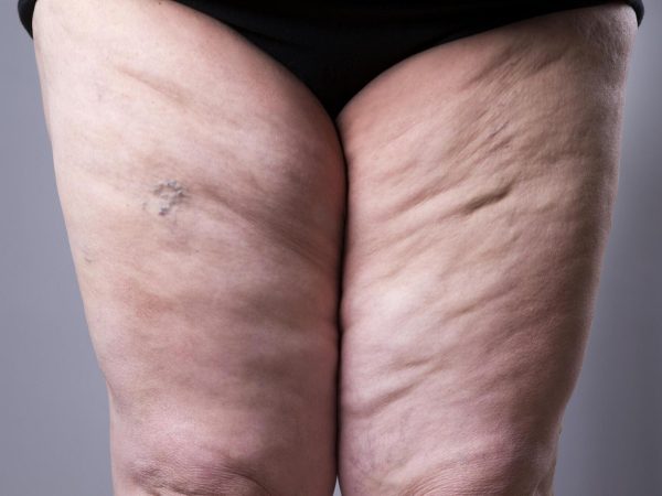
Lipedema is about more than genes or hormones. The disease is characterized by inflamed subcutaneous adipose tissue (SAT), tenderness, easy bruisability, and symmetric but frequently asymmetric fat hypertrophy. It affects almost 11% of adult women globally, and inheritance is seen in up to 60% of cases, indicating a strong genetic predisposition that acts in conjunction with hormonal and local tissue factors.
Here are some fundamental co-factors that influence course, severity, and treatment requirements.
Impaired Lymphatics
Lymphatic dysfunction decreases fluid clearance from tissues. As a consequence, suboptimal lymphatic drainage causes fluid accumulation in the interstitium resulting in edema, which compresses adipocytes and further exacerbates SAT expansion. Lymphatic capillaries and collecting vessels routinely shuttle protein-rich fluid away from tissue.
When they don’t, proteins and immune cells remain, altering tissue tension and encouraging fibrotic transformation of fat. This dysfunction puts you at risk of overt lymphedema. Once lymph flow becomes chronically impaired, the resulting swelling is more difficult to undo.
Lymphatic loss raises infection risk and delays healing in those legs. It’s not a part of clinical care, which should include lymphatic-focused options. Manual lymphatic drainage, compression therapy with graded garments, pneumatic pumps, and skin care to avoid infections are crucial elements.
Imaging such as lymphoscintigraphy can assist in evaluating flow and directing treatment options.
Vascular Fragility
Vascular fragility manifests as easy bruisability and predisposition to orthostatic edema. Delicate microvessels ooze fluid and blood elements into SAT, nourishing localized inflammation and discoloration. When the delivery of nutrients and oxygen to subcutaneous tissue is compromised, adipose cells can react by hypertrophying and attracting inflammatory cells.
Vascular disease can therefore exacerbate fat infiltration and aggravate pain. Poor microcirculation impedes drug delivery and healing, which can reduce the effectiveness of certain treatments. Things to monitor are easy bruising following minimal trauma, edematous ankles that fluctuate with standing and persistent discoloration or petechiae.
Tacking a few of these on helps clinicians identify a vascular element earlier.
Inflammation
Inflammatory markers increase in the tissue involved, immune cells invade the SAT and local cytokines alter fat cell behavior. This process accounts for stubborn pain, inflammation and diet, exercise or bariatric surgery resistance in so many patients.
Anti-inflammatory strategies can help.
-
Anti-inflammatory diet of real food, omega 3s, and low processed sugar.
-
Regular low-impact exercise to support circulation.
-
Weight-bearing compression during activity.
-
Pharmacologic options: NSAIDs short-term, targeted meds as advised.
-
Therapies include lymphatic drainage, shockwave, or targeted procedures to reduce inflammation.
Listing all contributing factors aids comprehensive care: genetics, hormones, lymphatic impairment, vascular fragility, chronic inflammation, biomechanical stress, and lifestyle factors such as activity level and diet.
Diagnostic Challenges
Diagnosing lipedema takes a thoughtful, systematic approach because it is frequently misdiagnosed as something else. Physicians have to parse overlapping symptoms like excess fat, swelling, and easy bruising while considering patient history and potential familial trends. Many patients wait years for a correct diagnosis because symptoms can appear non-specific and develop gradually.
Proper diagnosis mixes clinical criteria, specific imaging, and thorough history to differentiate lipedema from obesity, lymphedema, and other fat conditions.
Lipedema vs. Obesity and typical fat distribution. Obesity typically exhibits a more even fat gain throughout the body and responds to calorie restriction and exercise. Lipedema features disproportionate fat on the legs and sometimes arms, often painful, tender, and sparing of the feet.
About diagnostic conundrums, patients describe heaviness and stiffness that far exceed ordinary weight gain. Weight loss can reduce trunk fat but leaves limb fat largely unaffected, which should tip clinicians off to an alternate process.
Lipedema vs. Lymphedema and edema – diagnostic challenges. Lymphedema commonly presents with swelling including the feet and in the early stages with skin changes and pitting. Lipedema usually spares the feet, produces soft nodular tissue and bruises easily.
There can be secondary lymphatic dysfunction in advanced lipedema, muddying the waters. Differentiating primary lymphedema, chronic venous disease, and lipedema all require specific physical exam maneuvers and occasionally lymphoscintigraphy or ultrasound.
Typical misdiagnoses are saying lipedema is just obesity, that the pain is orthopedic and that swelling is “edema” without cause. Symptom overlap—pain, swelling, bruising, heaviness and impaired mobility—fuels these missteps.
Misdiagnosis results in treatments that range from strict dieting alone or unnecessary procedures to delaying symptom-focused care like compression, manual therapy, or liposuction referral when warranted.
Clinical tools and testing assist. Use standardized clinical criteria: symmetrical limb enlargement, sparing of hands and feet, tenderness, and easy bruising. Gather a thorough family history. Research shows as many as 60% of people with lipedema have a relative with the disorder.
Imaging, like ultrasound, MRI, or lymphoscintigraphy may reveal typical fat distributions or lymphatic modifications. Record symptom onset, response to weight loss, and functional limitation.
Emotional and pragmatic consequences count. The diagnostic process may be prolonged and exasperating, and patients commonly experience hopelessness, low self-esteem, and anxiety.
Coordinate care, validate patient reports, and set realistic steps. Rule out common causes, use imaging when needed, and involve specialists early.
A Personal Perspective
Lipedema is the silent guest that overstays its welcome. A lot of readers will recognize the gradual emergence of odd pockets of fat on the legs and arms in puberty or beyond, followed by years of frustration as dieting and exercise don’t affect the distribution.
This section shares why personal stories matter, how the condition affects daily life, and which small practical changes can help. Lipedema patients report varying symptoms and different times of onset. They may observe the first signs starting between 10 and 19, but then wait an average of 17 years to receive a diagnosis.
That delay matters. Mislabeling the issue as simple obesity or as lymphedema is common, and it delays targeted care. Sixty to eighty percent of cases are familial, so identifying relatives with similar leg or arm transformations can accelerate diagnosis. Others highlight particular genetic variants that increase risk, though genetics alone cannot account for why one individual is impacted while another is not.
It impacts quality of life in very tangible ways. Pain, easy bruising, and heaviness complicate walking, standing, or travel, while progressive fat deposits can further restrict movement as time goes on. These corporeal constraints transform labor and play decisions.
Self-image often suffers; many report feeling self-conscious or embarrassed, which can lead to social withdrawal and stress. Patient narratives aid physicians in seeing past statistics and body mass index metrics to understand how symptoms impact everyday life.
Opening detailed personal experiences makes better care and community. Include when symptoms started, what treatments you tried and what helped or exacerbated. Note triggers such as hormonal changes, stress, or weight gain.
Include measured details such as how far you can walk, what clothing fits, whether compression reduced pain, and how long surgical or manual therapies gave relief. These details lead others and assist scientists to identify general trends.
Practical self-help concepts from others’ lives can be beneficial. Compression garments can alleviate the pain and swelling for hours or days. Try different fabrics and pressures until you find a comfort level.
Light, weight-bearing exercise such as swimming or walking aids mobility without overburdening painful spots. Manual lymphatic drainage, trained massage, and skilled physiotherapy frequently help to diminish symptoms.
Diet shifts won’t cure cellulite but can reduce inflammation and aid vitality. Prioritize wholesome meals, sufficient protein, and minimal processed food selections. Keep track of what you attempt and the impact over weeks to cultivate a helpful personal list.
Conclusion
The signs are that hormones and genes play a definitive role in lipedema. Hormone shifts connect to symptom changes at puberty, pregnancy, and menopause. Genetic alterations appear in families and in analyses of specific gene variants. Other things like weight gain, inflammation, and lymph strain define how the condition appears and how quickly it progresses.
It is helpful to have a balanced perspective. Seek treatment that addresses hormones, genetics, and tissue. Easy moves such as targeted exercise, compression, and anti-inflammatory options can relieve symptoms. For a clear plan, find a lipedema knowledgeable clinician who provides tests and options that align with your life. Need assistance locating resources or crafting questions for your appointment? I can assist.
Frequently Asked Questions
What is lipedema and who does it affect?
Lipedema is a chronic condition that causes symmetrical fat accumulation, typically in the hips, thighs, and arms. It primarily impacts women and tends to emerge or exacerbate during hormonal events such as puberty, pregnancy, or menopause.
Are hormones the main cause of lipedema?
Hormones are a big but not a solo cause. Estrogen and other female hormones are associated with symptom onset and progression, particularly around life stages with hormonal fluctuations.
Is lipedema genetic?
Yes. Family patterns and studies show that there is a genetic component. Multiple genes probably contribute, making some people prone to getting it.
Is lipedema caused by both hormones and genetics?
Most evidence points to a combined effect. Genetic predisposition and hormonal triggers usually account for the timing and worsening of lipedema.
Can lifestyle changes reverse lipedema?
No. Diet and exercise assist in symptom management, inflammation reduction, and mobility improvement, but they seldom get rid of lipedema fat. Early care decelerates advancement and improves quality of life.
How is lipedema diagnosed?
Diagnosis is clinical, based on history and physical exam. Experts search for symmetrical fat, pain, easy bruising, and resistance to conventional weight loss. Imaging can aid to rule out other causes.
What treatment options exist for lipedema?
Treatment focuses on symptom control, including conservative care such as compression, manual lymphatic drainage, and exercise. Pain management is also important, along with surgical options like lipedema-specific liposuction for advanced cases. Early treatment enhances results.