Key Takeaways
-
Lipoedema tends to present with symptoms such as pain and is most commonly found in the legs and arms, whereas ‘stubborn fat’ can occur anywhere on the body and is not painful.
-
That’s why proper diagnosis with ultrasound is so important — lipoedema isn’t stubborn fat.
-
Ultrasound imaging provides a non-invasive method to distinguish fat varieties, evaluate tissue texture, and detect unique characteristics such as fluid accumulation and fibrosis strands, bolstering targeted treatment strategies.
-
We know that operator knowledge of ultrasound is essential for an accurate diagnosis, so we recommend seeking care from trained professionals to obtain a dependable diagnosis.
-
Dealing with overlapping signs and concurrent conditions necessitates an in-depth clinical evaluation, which involves taking a comprehensive history, conducting a physical examination, and consulting across disciplines.
-
To maximize efficacy, pair ultrasound insights with whole-body care approaches and get real about what non-surgical fat reduction can and cannot achieve.
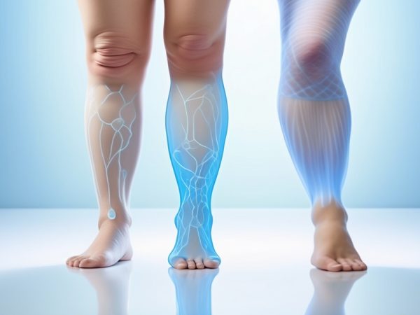
Lipoedema and ‘stubborn fat’ appear to be similar on the surface, but check out what ultrasound scans reveal. Lipoedema is a long-term disease in which fat accumulates in a fixed pattern in the legs and arms and frequently triggers pain and swelling. Stubborn fat, in comparison, is simply residual body fat that lingers post diet or exercise and doesn’t have the pattern. Ultrasound can reveal how lipoedema fat spreads beneath the skin and differs from normal fat. The scan aids physicians in differentiating lipoedema from typical fat and directing appropriate treatment. In the following, discover how ultrasound images provide a clean window into each, and find out why this imaging is important in actual treatment.
The Two Fats
Lipoedema and resistant fat are different in many respects, from cause to presentation to therapy. Although both consist of fat, they each have distinct symptoms and impact on the body.
-
Lipoedema is a long-term medical issue characterized by unnatural, symmetric fat deposits, typically in the legs and arms. It frequently hurts, swells and bruises easily, unlike stubborn fat which is painless and located in traditional storage areas such as the stomach and hips.
-
Lipoedema fat occurs mainly on the lower and upper limbs on both sides, with no effect on the hands or feet. Stubborn fat can appear anywhere but tends to be most apparent on the belly, thighs and occasionally the upper arms.
-
Pain and discomfort are typical with lipoedema. Stubborn fat does not create these symptoms.
-
Hitting the proper diagnosis is critical. Lipoedema needs a specialist, whereas persistent fat is often self-diagnosed or consulted with a GP.
Lipoedema
Lipoedema is a long-term fat metabolism disease. It’s deep subcutaneous fat, with special metabolic characteristics such as changed amino-acid and phospholipid production. Unlike regular gynoid fat, the lipoedema fat doesn’t shrink with a calorie deficit. It resists both diet and exercise. Cellulite and lipoedema are not the same: cellulite is a cosmetic issue at the hypodermal level, while lipoedema is a medical problem at a deeper layer. Lipoedema tends to cause an increased body fat percentage than those with comparable BMI without the condition.
It ranges from mild swelling and discomfort to more advanced stages, where mobility is impaired. Therapeutic decisions are stage dependent, with earlier phases being more responsive to conservative treatments such as compression and manual therapy. Late may require surgery.
There is a genetic element to lipoedema. The research points to genes, such as Bub1, as potential candidates. Family history may increase risk, making awareness and early intervention key.
You require specialist care. While these are not a cure, care such as lymphatic drainage, custom compression garments and specialized surgery can help manage symptoms better than typical weight loss methods.
Stubborn Fat
Stubborn fat is normal subcutaneous or visceral fat that fights diet and exercise. It can show up anywhere, but more frequently in the belly, thighs or hips.
So many of us are fed up with stubborn fat. It can impact confidence in a body-centric world. Unlike lipoedema, stubborn fat isn’t painful, but it can be upsetting.
Conventional liposuction and body sculpting typically do the trick for hard-core fat, particularly in individuals close to their target weight.
The Ultrasound Lens
Ultrasound helps tell the real story between lipoedema and persistent fat. By pulsing sound waves into the tissue, it reveals what’s happening beneath the surface—no incisions, no speculation. Physicians depend on these images to examine fat texture, thickness, fluid and others. This directs diagnosis and treatment, from non-invasive fat removal to ultrasound-assisted liposuction.
Tissue Texture
Lipoedema and hard to lose fat appear differently on ultrasound. Lipoedema tissue can feel grainy or uneven, whereas stubborn fat is smoother, with more consistent fat layers.
Doctors notice the brightness or darkness of tissues on the scan—this is referred to as echogenicity. Lipoedema appears heterogeneously bright, suggesting fibrous and inflamed regions. Stubborn fat tends to appear more flat and less dimpled. Texture analysis really does count. It supports the early detection of lipoedema and prevents it from being misdiagnosed as hard-to-lose fat, ensuring individuals receive appropriate treatment.
Fat Lobules
Fat lobules are little fat pockets, and their shape counts. On ultrasound, lipoedema lobules are larger, more spread out and less structured than in stubborn fat. Normal hard-to-lose fat lobules are typically small and densely packed.
This distinction assists physicians in determining whether they’re dealing with a fat disorder or just plain old stubborn fat. It impacts treatment decisions as well. For instance, ultrasound-assisted liposuction can shatter bigger, lumpy lobules more effectively than regular techniques. Understanding the lobule arrangement aids in managing patient expectations.
Skin Thickness
Lipoedema usually has skin that’s thicker than resistant fat. Ultrasound makes it simple to quantify. Occasionally, a couple of spare millimeters can indicate lipoedema – particularly in the legs and arms.
Thicker skin can imply greater swelling or chronic underlying change. This has an impact on what treatments are most effective, and how long it may take to recover. Monitoring skin thickness over time reveals whether treatments, such as ultrasound cavitation, are indeed assisting.
Fluid Pockets
Fluid pockets, or edema, are simple to identify on ultrasound with lipoedema. These appear as shadowy areas between fat lobules.
Swelling and soreness results from fluid accumulation. For lipoedema, it frequently involves flushing this additional fluid. Ultrasound assists physicians select therapies that address fluid, such as manual drainage, and monitor its efficacy.
Spotting fluid pockets early can really make a difference in comfort and outcome.
Fibrotic Bands
Fibrotic bands appear as dense, bright lines on the scan. These are rigid, rope-like connective tissues occurring more in lipoedema.
They alter fat deposition and treatment absorption. For instance, tight bands can render fat tougher to extract, even using ultrasound-assisted liposuction. Following these bands over time assists physicians in modifying plans and managing symptoms of them long-term.
Diagnostic Nuances
Ultrasound provides unhindered real-time look at tissue under the skin but is not a panacea. The specifics detected by the scan are sometimes difficult to interpret, and what is noticed varies significantly among practitioners. The distinctions between lipoedema and hard-to-lose fat are not always clear, and at times they present simultaneously or intermix with other conditions.
Operator Skill
Talent counts. Ultrasound images require an experienced hand to obtain and interpret them correctly. Veteran technicians recognize these implicate indications, such as locally thickened fat layers or small fluid collections, that more novice eyes might overlook. They know where to look and how far down, and that can alter a diagnosis.
Regular practice is crucial. Not all clinicians receive the same training or education in ultrasound. Training gaps can result in mistakes—overlooking the ache from hypoxic fat in lipoedema, or mistaking gravity-induced swelling at day’s end for pathology. Operator expertise informs patient results, from initial imaging to therapy design.
Overlapping Signs
-
Both may demonstrate hypertrophy of the subcutaneous fat to the extremities, particularly the lower body.
-
There can be mild swelling at the end of the day.
-
Won’t reduce in swelling with leg elevation or sleep in hard core fat
-
Skin may appear normal, without apparent textural changes in both
-
Patients can have a higher BMI in either group
The detailed health history sifts it all. Family history, symptom onset and progression all indicate lipoedema or hard-to-lose fat. A complete examination—palpating, pressing, observing how the swelling behaves—provides more hints. Folks often believe that any sluggish fat loss equates to lipoedema, or that all leg fullness is stubborn fat, but these are not trustworthy indicators.
Coexisting Conditions
-
Chronic venous insufficiency
-
Obesity
-
Lipo-lymphedema
-
Heart or kidney disease
These can muddy the waters further. When lipoedema joins forces with other problems such as obesity or lymphedema, symptoms intertwine and become more intricate. Swelling doesn’t just disappear overnight. Pain escalates and the likelihood of skin changes increases. It requires a team of specialists—vascular, lymphatic, dermatology—to untangle and address the underlying causes. Only a full checkup covers all bases.
Beyond The Image
Lipoedema and ‘stubborn fat’ can look very similar but behave completely different below the surface. Ultrasound provides an intimate glimpse, yet it captures a mere fraction of the whole narrative. Living with lipoedema isn’t just about what meets the eye. It’s about pain and change and what it means to feel at home in your body. Patient stories, physical exams, and teamwork in care all count in ways images can’t always capture.
Patient Story
Marta, a 38-year-old teacher, had lived for years with swelling and pain in her legs. She experimented with several different diets and exercised regularly, but nothing changed the way her legs felt or looked. Friends assumed it was simply excess weight, but her pain multiplied every year. Doctors overlooked lipoedema for ages. When she finally sought assistance, an ultrasound detected thick layers of fat and fluid under her skin, not “stubborn fat.” Treatment provided more than reduced pain—it returned daily comfort to her life and allowed her to walk unabashed. Marta now shares her story to assist others in recognizing that these diseases run deeper than the skin.
Too many, like Marta, still feel ashamed or alone. For a few, cellulite is par for the course, however for others it’s a source of upset and anxiety. By telling these stories, it helps people realize lipoedema and cellulite are legitimate physiological issues—not just aesthetic imperfections.
Physical Exam
A good physical exam for lipoedema examines more than girth. Doctors will examine skin texture, palpate for tenderness, and observe fat distribution on legs and arms. They might squeeze the skin to check for pain, or press to examine swelling. This practical check aids in detecting the dense, bread-sponge-like fat of lipoedema, which is distinct from standard fat or cellulite.
Ultrasound adds nuance but cannot substitute touch and eyes. Physical exams can reveal changes over time and guide a personalized treatment plan — not just what the scan shows.
A Holistic Diagnosis
Placing ultrasound findings alongside a patient’s history and comprehensive exam yields improved diagnoses. Lipoedema, cellulite and normal fat all appear distinctive. Ultrasound can detect thick fat layers, water retention, or fibrosis, but it doesn’t explain the entire tale.
Lifestyle, family history and daily symptoms count. Certain individuals experience both cellulite and lipoedema, with each causing painful swelling in distinct ways. Doctors collaborate—interview, scan, and exam—to identify the correct diagnosis and strategy.
Empathy in Practice
Personal stories shape how care is given.
Listening to patients builds trust.
Empathy helps both sides understand the struggles and hopes.
It results in superior care for actual humans, not merely their photographs.
Ultrasound Therapies
Ultrasound therapies provide non-invasive fat loss and contouring treatments. They blast fat cells in specific areas with sound waves, so folks can notice a difference without surgery or downtime.
Cavitation
Cavitation utilizes low-frequency ultrasound waves to form bubbles in the fluid surrounding fat cells. These bubbles pop and explode the fat cells. Following this, the body evacuates the debris on its own timetable.
What makes this approach ideal is that it’s most effective on small, pesky areas – under the chin, the knees, your love handles. Although they can notice a change after only one visit, a set of 1 – 3 sessions is generally necessary. Each session typically extends from 30 to 60 minutes. Most return to their regular day shortly after.
Unlike surgical liposuction, cavitation has no incisions or stitches, so there’s no downtime or scarring. It’s safe in the hands of trained personnel. Side effects tend to be mild, such as swelling or stiffness, and resolve within a few weeks.
HIFU
HIFU, or High-Intensity Focused Ultrasound, directs focused sound energy deep beneath the skin. This heat simultaneously damages fat cells and tightens skin. The body then proceeds to gradually dispose of the fat cells over the course of weeks.
HIFU can assist sculpt the body and tighten lax skin. It’s most useful for individuals with mild to moderate fat deposits and some skin laxity. People with more fat or loose skin might require additional treatments or extended sessions.
Benefits and Ideal Candidates
Ultrasound therapies allow patients to target little fat pockets that refuse to disappear despite diet or exercise. They are non-invasive and don’t require significant recovery time. Full results come in 6 to 12 weeks, though some require additional visits. Research indicates these therapies do not adversely affect insulin levels. Best for those near their target size looking to dial in their silhouette.
Treatment Realities
Ultrasound and similar non-invasive fat reduction treatments are popular for those seeking to dial into specific areas of fat, such as lipoedema or stubborn fat. These treatments are cozy, pain-free, with minimal downtime. They have their pros and cons, so definitely consider each aspect prior to jumping in. Below is a checklist to consider: check the provider’s experience, ask about clinical evidence, understand the treatment plan, review cost, discuss possible side effects, and set realistic expectations for results.
Efficacy
Clinical studies demonstrate ultrasound fat reduction can assist body contouring, cellulite reduction, and skin tightening for individuals near their ideal weight. Results are often incremental, with shifts becoming more apparent over months and multiple sessions.
Treatment outcomes vary based on body type and lifestyle. Folk that eat healthy, work out and keep a stable weight tend to do better. It’s patient commitment, first of all—skipping sessions or ignoring aftercare can hamper results.
Follow ups like ultrasound or simple measurements monitor fat loss and skin changes over time. Such input is valuable to both patients and providers when readjusting a plan.
Safety
|
Treatment |
Invasiveness |
Pain Level |
Downtime |
Risks |
|---|---|---|---|---|
|
Ultrasound |
Non-invasive |
Low |
Minimal |
Stiffness, swelling |
|
Laser Lipo |
Minimally |
Low-Mod |
Few days |
Burns, infection |
|
Surgical Lipo |
Invasive |
High |
Weeks |
Bleeding, scarring |
Potential hazards for ultrasonic are mild swelling or stiffness, typically dissipating within weeks. Very rarely, infection may happen if after care is not adhered to. Patients need to be informed consent, have clear education about what to expect. In the hands of experienced clinicians, these dangers can be minimized with appropriate dosing and careful observation of patients.
Limitations
Ultrasound treatments are not for morbid obesity or significant weight loss. They’re best for sculpting small trouble areas. Anatomy counts—fat thickness, skin quality, genetics all play a role in the results. Others might require a combination treatments, such as radiofrequency or LED for optimal outcomes.
Research continues to refine these treatments, but for the time being, outcomes are inconsistent and may not rival surgery.
Realistic Expectations
Treatments are 30-60 minutes, cost $150 – $3,500 per session and typically come in a series. Results can be subtle and slow so patience is key.
Conclusion
Ultrasound aids in mapping actual markers that differentiate lipoedema from standard stubborn fat. The scan provides a detailed view of tissue layers, edema, and the distribution of fat. It can reveal thick skin, increased fluid, and unusual pockets not visible with normal fat. Physicians can detect symptoms earlier and initiate appropriate programs sooner. This aids people discover best ways to address symptoms, from exercise to physician assistance. Knowing what appears on screen removes some of the mystery. For those who live with pain, swelling or weight that won’t quit, it could be worth querying a doctor about an ultrasound. When the facts are clear, it’s easier to mold a clever strategy and feel in control of your treatment.
Frequently Asked Questions
What is the difference between lipoedema and stubborn fat?
Lipoedema is a disease of abnormal fat accumulation, primarily in the legs and arms. Stubborn fat is just your garden variety fat that won’t budge very easily with diet and exercise. Lipoedema generally needs to be diagnosed by a doctor.
How does ultrasound help distinguish lipoedema from stubborn fat?
Ultrasound can show tissue differences. In lipoedema, ultrasound reveals pathological fat architecture and edema, whereas in stubborn fat, it’s homogenous adipose tissue. It assists physicians in making a more precise diagnosis.
Can ultrasound alone diagnose lipoedema?
No, ultrasound is a useful means, but not the exclusive one. Diagnosis should entail a physical exam, med history, and symptoms review by a knowledgeable healthcare provider.
Is lipoedema caused by poor lifestyle choices?
No, lipoedema is not lifestyle or diet related. It’s a chronic disorder related to genetics or hormones. Good habits might contribute to general wellness but don’t typically ‘fix’ lipoedema.
Are ultrasound therapies effective for treating lipoedema?
Certain ultrasound treatments can alleviate swelling and pain for lipoedema sufferers. These treatments are often a piece of a broader treatment regimen, not a cure.
What treatment options are available for lipoedema?
Treatment typically involves compression therapy, manual lymphatic drainage, exercise, and occasionally surgery. A healthcare provider can suggest the optimal choices.
Why is early diagnosis of lipoedema important?
Early diagnosis is key to managing symptoms, halting progression, and enhancing quality of life. It lowers the risk of complications, including pain and difficulty moving.