Key Takeaways
-
Lipedema is a unique, long-term fat condition that leads to irregular, typically symmetrical fat deposits in the lower body and predominantly impacts females. Early detection mitigates misdiagnosis and issues.
-
Diet and exercise alone rarely solve lipedema fat deposits, and management centers around a blend of conservative therapies, specialized nutrition, and when necessary, surgical excision of sick fat.
-
Signs indicate lipedema’s core issues are with fat tissue abnormalities, genetics, and hormones. Certain research reveals immune response and inflammation without diagnostic autoimmune markers. Thus, its categorization is still being researched.
-
There are no diagnostic lab tests for lipedema, so diagnosis relies on clinical history, physical exam findings such as nodular fat and non-pitting edema and imaging when indicated.
-
Because patients endure stigma, frustration, and emotional distress from misdiagnosis and futile weight-loss attempts, patient-centered care and greater public and professional awareness are critical.
-
Clinicians should adopt a stepwise diagnostic approach, rule out other conditions, record symptom progression, and provide personalized treatment plans including compression, manual therapies, exercise, and referral for specialist care when indicated.
Lipedema is a chronic fat disorder that primarily impacts the legs and arms and isn’t an autoimmune disease. It involves abnormal fat growth, pain, easy bruising, and swelling that progressively worsens over time.
Research connects hormonal and genetic factors and lymphatic changes to classic autoimmune causes. A clear diagnosis is important for treatment decisions such as compression, manual therapy, and targeted surgery.
The main body will go into signs, tests, and care options.
Defining Lipedema
Lipedema is a long-term condition that involves unusual, symmetrical fat accumulation below the skin, typically in the lower regions of the body. It usually manifests in women during puberty or other hormonal changes like pregnancy or menopause. This fat is nodular and often painful, forming a distribution that spares the hands, feet and trunk while establishing an obvious lower-body dominance.
This results in a significant upper to lower body disproportion, with waist to hip ratios frequently less than one. The gynoid distribution, which includes the lower abdomen, hips, buttocks, thighs and lower legs, is the predominant pattern.
The Misconception
A lot of folks think lipedema is just eating too much or not exercising. That view ignores clinical features: nodular, pearl-sized fat deposits, symmetric involvement, and pain in affected tissue. It’s easy for clinicians and patients to confuse lipedema with regular old-fashioned obesity as both present with excess fat.
Lipedema fat defies typical weight-loss strategies. Diet or exercise alone generally decrease non-lipedema fat and leave lipedema tissue mostly intact. Calling lipedema ‘weight gain’ postpones proper diagnosis and treatment.
Women may attempt one diet or fitness program after another to no avail and then feel at fault for their lack of success. This delay can allow progression. Stage 1 skin may feel smooth over nodular fat. Stage 2 shows indentations over larger masses. Stage 3 develops folds and deforming lobules that limit movement.
Public awareness is essential to minimize stigma and promote prompt medical evaluation. Better recognition by primary care providers and fitness professionals gets patients the right referrals and support earlier.
The Reality
Lipedema is a unique condition, with delineated symptoms and a definable trajectory. The disease is predominantly limited to the lower body and almost never involves the hands or feet. Arms are affected in approximately 80% of cases, but lower-body distribution prevails.
Swelling and increases in tissue fluid and fat can distort limbs, making ambulation and personal care more difficult as time goes by. Diet and exercise are healthy but don’t get rid of lipedema fat deposits.
Conservative measures, including compression, manual lymphatic techniques, and tailored physical therapy, improve symptoms and function but frequently don’t eliminate the abnormal fat itself. When indicated, surgical options such as lipedema-specialized liposuction can reduce tissue volume.
Both genetics and hormones are implicated. Approximately 15% of patients have a family history, and the onset around hormonal changes suggests an endocrine component. Diagnosis is largely clinical, relying on history and physical exam rather than a single lab test.
Earlier recognition helps with management and prevents disability.
The Core Debate
Lipedema sits at the center of a clinical disagreement: is it primarily a disorder of abnormal adipose tissue or a condition driven by immune dysfunction? The response influences how clinicians screen, treat, and prioritize research. Because definitions influence trials, reimbursement, and patient care, the stakes are pragmatic as much as scientific.
1. Fat Disorder Evidence
Several imaging and histologic studies describe enlarged, fibrotic adipocytes and altered extracellular matrix in affected tissue. Biopsies demonstrate greater fat lobulation and connective tissue septa than typical obesity. These structural distinctions are why fat in lipedema resists calorie restriction and exercise. Patients lose everywhere else, but lower-body fat stays.
Family pedigrees are well delineated. Almost all of my patients have female family members with the same leg or arm enlargement starting at puberty or following hormonal events, confirming the hereditary component. Hormone connections, especially to estrogen, are theorized since onset frequently occurs alongside puberty, pregnancy, or menopause shifts.
The centrality of fat tissue change is supported by clinical progression: slow, symmetric enlargement of fat compartments, nodular subcutaneous tissue, and a chronic course that can impair mobility and daily function. This can spiral into severe disability and psychosocial distress if misdiagnosed as ‘just’ obesity.
2. Autoimmune Arguments
A few studies report low-grade inflammation in lipedema tissue, such as elevated immune cell infiltration and pro-inflammatory cytokines. Patients occasionally complain of tenderness, burning, and lingering pain, symptoms that can mirror immune activation as opposed to just mechanical fat expansion.
Clinically, lipedema overlaps with autoimmune diseases in subsets of patients, indicating common mechanisms in at least some instances. Different reports and causation are not determined. Immune markers specific to classic autoimmune diseases are typically absent in lipedema cohorts.
Most importantly, no single, consistent autoimmune biomarker characterizes lipedema. That absence of conclusive markers renders it too early to classify lipedema as an autoimmune disease. Immune mechanisms could amplify symptoms and progression in certain patients.
3. Clinical Distinctions
Lipedema usually displays symmetric fat deposition involving hips, thighs, and upper arms with sparing of hands and feet. This contrasts with lymphedema, where swelling can be asymmetric and involve distal extremities.
Easy bruising and pain is common and can be severe, which distinguishes lipedema from mild obesity. Non-pitting edema and nodular fat on exam differentiate it further. These characteristics, along with the disease’s chronic, progressive nature and its common onset in girls at puberty, support GP as a unique clinical entity.
A table contrasting lipedema, obesity, and lymphedema clarifies differences in distribution, edema type, pain, and response to weight loss.
4. Diagnostic Markers
Physical exam reveals nodular subcutaneous tissue, tenderness, and non-pitting edema. Ultrasound and other imaging can identify hyperechoic septa and tissue densification.
There is no blood test that consistently diagnoses lipedema. Clinical history, symptom patterns, and directed imaging continue to be fundamental. With such a high misdiagnosis rate, low awareness among specialists, and the significant treatment differences, proper classification is essential for the right treatment and better outcomes.
5. Expert Consensus
Guidelines more and more define lipedema as a specialized fat disorder, not classic obesity. Although many specialists still pursue immune connections, the diagnostic standards are shifting as data accumulates. Broader awareness and more transparent standards will enhance treatment and limit psycho-social damage.
Symptom Profile
Lipedema has a unique symptom profile that distinguishes it from other edema disorders. Lipedema primarily impacts the distribution and storage of subcutaneous fat, generating bilateral, symmetrical enlargement of the lower extremities with sparing of the feet. Symptoms progress and frequently worsen with hormonal fluctuations or general weight gain.
-
Disproportionate lower body fat (hips, thighs, buttocks)
-
Leg heaviness and pain on touch
-
Symmetrical swelling of both legs
-
Easy bruising and skin sensitivity
-
Soft, rubbery subcutaneous tissue and nodularity
-
Preservation of normal feet appearance
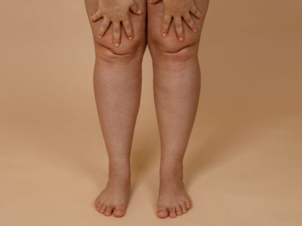
Physical Signs
The most obvious symptom is symmetrical swelling of the legs, typically beginning at the hips or upper thighs and progressing downward. Enlargement is bilateral, and in numerous patients the arms present similar though less pronounced alterations.
This symmetrical alteration represents distributed, symmetrical fat tissue dysfunction and adipocyte hyperplasia rather than localized fluid accumulation. Tissue feel is important in exam: affected tissue is soft and rubbery, with small, evenly spaced nodules in early stages and larger, uneven nodes in later stages.
Stage 1 skin is smooth with thick subcutaneous tissue and small nodes. By stage 2, the skin may take on an “orange peel” texture and palpable, irregular subcutaneous nodes appear. Skin is hypersensitive and easily bruised. Tenderness on pressure is frequent.
Feet are spared despite swelling and Stemmer’s sign is negative, which assists in differentiating lipedema from lymphedema. Neurological testing reveals tenderness to touch but not frank motor or sensory loss.
Progression may result in lipolymphedema, in which lymphatic load is augmented secondary to fat changes. Without care, this may progress toward frank lymphedema and worse.
Emotional Impact
A lot of patients endure psychological trauma because lipedema is misdiagnosed as straightforward obesity. This mislabeling brings social stigma and self-blame.
Frustrating, failed weight-loss attempts compound emotional distress and can engender a sense of despair in patients who adhere to diet and exercise only to witness stubborn fat remaining. They’re at increased risk for anxiety and depression associated with changed body image and longterm pain.
Concerns regarding future mobility, the financial costs of treatment, and repeated clinical encounters factor in.
-
Frustrated that diet and exercise do not shrink affected areas.
-
Feeling isolated because appearance does not match effort.
-
Confidence lost to chronic pain and mobility bounds.
-
Worry about long-term health risks such as hypertension or diabetes.
Diagnostic Pathways
Diagnosing lipedema follows a stepwise approach rooted in clinical assessment, targeted history taking, exclusion of other disorders, and selective imaging when needed. The process centers on careful observation of fat distribution and symptoms, with tests used to rule out lymphedema or vascular disease rather than to confirm lipedema alone.
Clinical Evaluation
Begin with a directed patient history noting symptom onset, familial incidence and flare initiators like puberty, pregnancy or weight fluctuations. Most patients describe slow, symmetric leg swelling with tenderness, frequent bruising and recalcitrant fat despite diet and exercise.
Record length and course of symptoms and observe if symptoms intensify with orthostasis or menses. Physical exam should evaluate fat distribution, consistence, and dolority. Lipedema usually causes soft, nodular subcutaneous fat with an ankle cuff and foot sparing.
Stemmer’s sign is negative. Examine for symmetry of the lower limbs and for pressure pain. Differentiate from obesity by disproportionate regional fat that resists weight loss and from lymphedema by the lack of positive Stemmer’s sign and associated toe sparing.
Employ published exam pathways where possible, which include measurements of limb circumference at set points and photographic records. Note skin changes, presence of pitting edema, and any limitations in mobility. Repeated measures over time assist in demonstrating progression and response to conservative care.
Diagnostic Challenges
No one lab test confirms lipedema. A recently reported biomarker holds promise to distinguish lipedema from other lymphatic disorders but is not yet widely available in routine practice. This absence of a conclusive blood test leaves clinicians depending on pattern recognition and elimination.
Symptoms often mimic obesity and lymphedema, leading to misdiagnosis. Lipedema, on the other hand, is rare and under-recognized, with many patients misdiagnosed or diagnosed late, which can result in worse patient outcomes.
If management is delayed, it can result in secondary lymphatic damage, which makes the clinical picture more similar to lymphedema. Imaging assists when the exam is ambiguous. Technetium-99m human serum albumin lymphoscintigraphy can evaluate lymph flow and detect the presence of lymphedema.
3D CT Angiography can reveal subcutaneous fat and exclude vascular causes. Use imaging sparingly to rule out alternative disorders rather than to confirm lipedema.
Education and awareness is key. To diagnose, educate clinicians to inquire about certain history items, conduct structured exams, and consider lipedema when symmetrical leg enlargement with tenderness and a negative Stemmer’s sign occur.
The sooner you diagnose and treat, the better the outcomes and the more limited the progression.
Management Strategies
Managing lipedema involves a layered approach that combines daily self-care, non-surgical treatments, and surgery when appropriate. Management strategies need to be individualized, considering disease stage, symptom burden, and personal goals. There is an emphasis on early intervention to decelerate progression and maintain function.
Conservative Care
-
Compression garments combat swelling and pain through tissue support and enhanced fluid return. Proper fit counts. Patients need to collaborate with trained fitters to choose garments with a metric-measured compression and replace them when elasticity diminishes. Several wear compression daily during activity and at night in a few cases.
-
Manual lymphatic drainage and specialized massage techniques assist in moving interstitial fluid and can reduce pain and discoloration. Certified lymphatic therapists and lipedema-sensitive methods are better than generic massage. Some patients supplement with measures like pneumatic compression devices at home for additional impact.
-
Low-impact exercise maintains flexibility, increases strength, and assists with pain and fatigue. Walking, swimming, cycling, and water-based classes minimize stress on the joints. One small study observed less pain and bruising when compression was combined with exercise. Exercise regimens need to be slow and cautious under the guidance of physical therapists who understand lipedema and are adapted to either fatigue or joint restrictions.
-
Nutritional advice is symptom management and overall health, not shrinking stubborn lipedema fat. Lipedema fat frequently resists diet, exercise, and even bariatric surgery. Therefore, nutrition is more focused on reducing inflammation, managing weight where appropriate, and boosting energy. Dietitians may suggest anti-inflammatory templates, real-world meal plans, and micronutrient audits during hormonal shifts such as pregnancy or menopause.
Other conservative measures include skin care to avoid breakdown, targeted supplements where evidence supports, stress management to reduce pain sensitization, and mental health support for chronic symptoms like brain fog and fatigue. Most patients tell me that it’s the daily discipline—consistent compression, exercise, and diligent self-care—that makes treatment effective over time.
Surgical Options
-
Liposuction, conducted with specialized techniques modified for lipedema, extracts the sick fat and may alleviate pain, swelling, and mobility restrictions. A 2023 study found quality of life enhancements following liposuction for numerous patients.
-
Surgery is typically reserved for after sufficient conservative care and when symptoms are considerably life-impacting. It’s not a front-line treatment, and it shines when integrated into a strategy.
-
Complications range from bleeding, infection, contour irregularities, and lymphatic damage. Potentially permanent reductions in size and pain depend on the technique, severity of disease, and post-operative care.
-
Selecting seasoned surgeons with experience in lipedema is key. Find clinicians that utilize metric outcome tracking, have peer-reviewed results, and work with therapists for pre and post-op rehab.
Where evidence is sparse, CoolSculpting, for instance, has few dedicated studies. Patients need to consider options and talk through expectations. Early, personalized treatment enhances quality of life and function.
A New Perspective
That lipedema requires a more precise definition that is based on recent research and patient testimonies. It is a heritable disorder of fat metabolism that results in symmetrical fat growth, frequently on the legs, characterized by an increase in adipocyte proliferation and abnormal fat storage. This distribution often spares the feet, is symmetrical, and demonstrates a negative Stemmer’s sign, which aids in differentiation from lymphedema.
The tissue is frequently painful to palpation and can induce persistent soreness and discomfort that permeate daily life and motion. Women frequently complain of being ‘thick in the legs,’ an alteration that strikes at identity and self-image in a culture that worships thinness.
Redefinition needs to get away from considering lipedema as just excess weight. Weight stigma causes most clinicians to overlook or downplay the disease, delaying treatment. A more precise nosology would separate lipedema from general obesity and autoimmune disorders by focusing on its unique pathophysiology, which includes altered lipid metabolism, symmetrical fat hyperplasia, and characteristic clinical signs.
It would alter research priorities, clinical guidelines, and insurance coverage decisions, and it would reduce the threshold for impacted individuals to access specialist care and surgery when indicated.
Research should continue on cause and treatment. Genetic, hormonal, and microvascular/inflammatory research could explain the initiation and progression of the condition. Trials should test diets, medications, and procedures with meaningful outcome measures such as pain, function, quality of life, and tissue change by imaging.
For instance, a ketogenic-style diet modified for lipedema and personal variation is promising for certain patients in minimizing inflammation and optimizing body composition. It requires rigorous research and well-defined recommendations on safety and sustainability.
Mindful medicine is about caring for both body and mind. Providers need to screen for pain, mobility restrictions, and psychological effects of disfigurement and social stigma. Conservative measures, such as compression, specific exercise, and manual therapies, pair with adjunctive approaches like vibration therapy, cupping, and Epsom salt baths to alleviate symptoms in certain individuals.
When surgery is on the table, shared decision-making should encompass realistic outcomes and post-operative care as well. Education for clinicians and patients is essential, as many cases are missed or undertreated because the awareness is low.
Public and professional awareness has to increase to make a difference. Primary care training modules, more transparent diagnostic criteria, and community resources can reduce diagnostic delays. Greater consciousness will reduce stigma, link patients to proven treatments, and nudge funders toward required research.
Conclusion
Lipedema sits between two perspectives. Research is showing it as a disorder of fat and fluid with distinct inflammation and tissue changes. It does not fit classic autoimmune disease by current lab tests and criteria. Yet immune cells are involved in the tissue destruction and pain. Clinicians use symptom checks, imaging, and patient history to guide treatment. Treatment combines weight management, exercise, compression, manual therapy, and surgery for others. A consistent routine and care that works for them helps a lot of people.
As a personal next step, consult a clinician with lipedema experience. Inquire about imaging, conservative care, and referral for lymphatic or surgical interventions.
Frequently Asked Questions
Is lipedema an autoimmune disease?
No. Lipedema is not an autoimmune disease. It’s a chronic fat disorder with vascular and lymphatic involvement. Immune research is underway.
Is lipedema the same as lymphedema?
No. Lymphedema is fluid buildup from lymphatic damage. Symmetrical painful fat accumulation is lipedema. They can occur together and are referred to as lipo-lymphedema, but they are distinct.
What causes lipedema?
We don’t know the cause. Hormones and genetics seem to be the big players. Inflammation and microvascular dysfunction may play a role, but a singular definitive cause has not been identified.
How is lipedema diagnosed?
Clinical diagnosis. Providers utilize history, physical exam, and exclude obesity and lymphedema. Imaging such as ultrasound or lymphoscintigraphy can assist but are not always necessary.
Can weight loss cure lipedema?
No. Diet and exercise can optimize overall health and reduce some edema, but they don’t typically eliminate lipedema fat. Specialized therapies and surgical intervention can provide permanent contour and symptom relief.
What are common treatments for lipedema?
Treatments range from conservative care, including compression, manual lymphatic drainage, and exercise, to surgical treatments such as lipedema-targeted liposuction. Pain management, along with psychosocial support, assists quality of life.
Should I see a specialist for suspected lipedema?
Yes. See a clinician experienced in lymphatic or adipose disorders, such as a vascular specialist, lymphologist, or plastic surgeon familiar with lipedema. Early evaluation improves management options.